


6 Bio-hybrid Tooth
- An Implant That Restores Natural Tooth Function

Ph.D. in Science from Kyushu University Graduate School. Has held positions including Professor at Tokyo University of Science, Team Leader at RIKEN BDR, and others. Currently Visiting Professor at Tokyo Dental College and Visiting Senior Scientist at RIKEN. Founded OrganTech in 2008 and became Chairman in 2024. View full profile >
When humans are born, the cells composing the body possess finite lifespans and are continuously replaced by newly generated cells. The human body consists of more than 200 distinct cell types, organized into hierarchical lineages composed of stem cells, progenitor cells, and differentiated cells, each giving rise to cells with predetermined fates. Even when tissues are subjected to disease or injury, this stem cell system functions to restore the original structure through endogenous regenerative mechanisms.
Various stem cell populations are also present in teeth. It has been well established that stem cells reside within the dental pulp, and that stem cells capable of generating the periodontal ligament and alveolar bone exist within the periodontal tissues. These periodontal ligament stem cells are reported to exhibit remarkable plasticity, differentiating into mineralized tissues such as bone or into ligamentous structures depending on local environmental cues (Chapter 1), although their precise regulatory mechanisms remain incompletely understood.
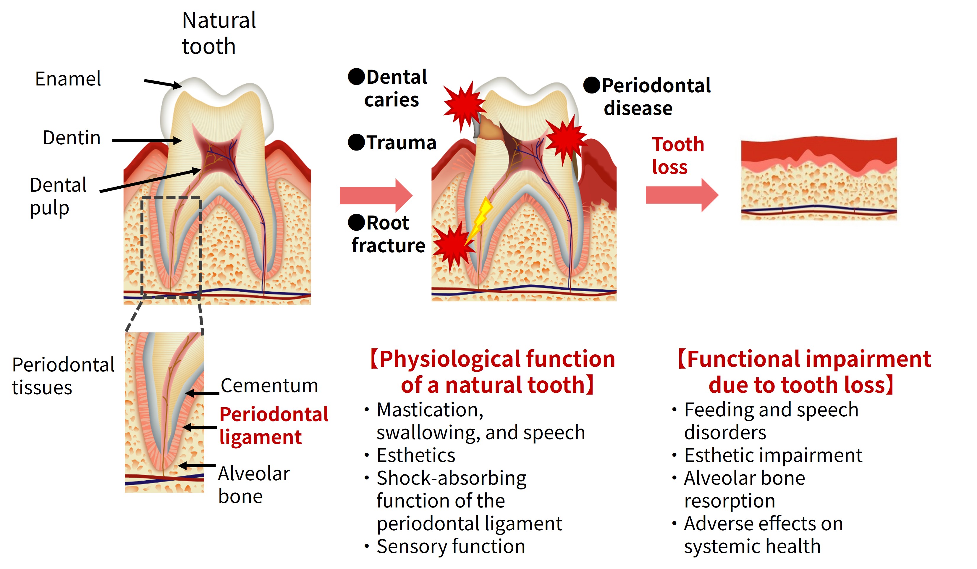
Tooth loss primarily results from periodontal disease, dental caries, or tooth fracture, leading to spontaneous tooth loss or extraction (Figure 1). In periodontal disease, bacterial infection destroys the periodontal ligament, resulting in tooth exfoliation. In contrast, during extraction due to caries or fracture, the periodontal ligament often remains biologically intact despite removal of the tooth. At the time of extraction, the ligament becomes torn and remains adherent both to the extracted tooth surface and to the extraction socket within the alveolar bone. It was therefore reasonable to hypothesize that periodontal ligament stem cells persist within these remaining tissues.
“This could be utilized.”
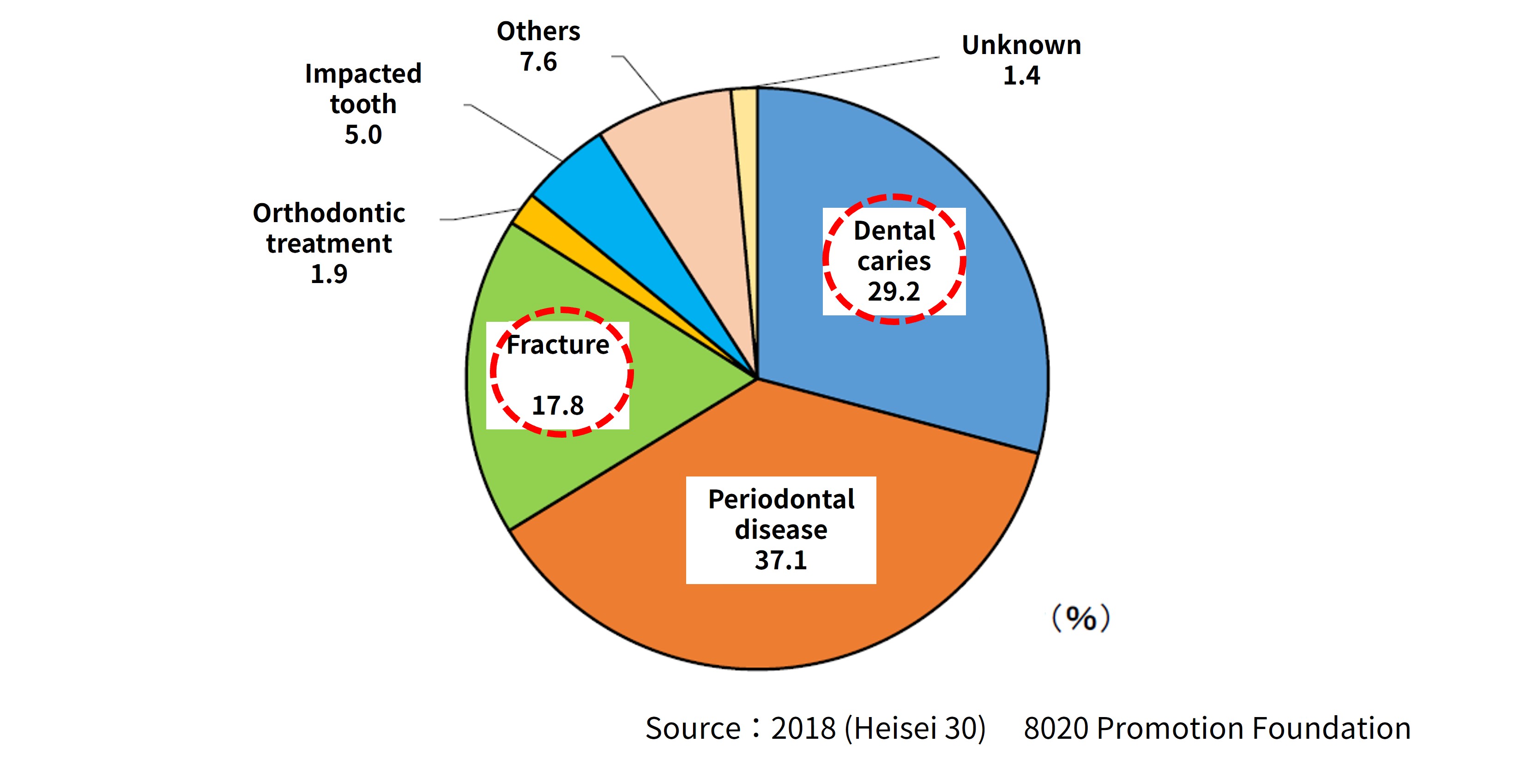
Isolation and culture of Periodontal ligament stem cells would require compliance with regenerative medicine regulations, involving substantial time and cost. However, tissues naturally remaining at the extraction site are not subject to such regulatory constraints. We therefore defined cases involving caries or fracture—where native periodontal ligament tissue can be utilized—as first-generation therapy, whereas conditions such as periodontal disease, in which healthy ligament tissue cannot be obtained and cell culture becomes necessary, were categorized as second-generation therapy. Our research began with the first generation. Epidemiological statistics indicate that approximately 48% of tooth loss cases correspond to first-generation indications (caries and fracture), while periodontal disease accounts for 37.1% (8020 Promotion Foundation, 2018; Figure 2).
A fundamental question then arose: how could the torn Periodontal ligament be integrated with an HA-coated implant surface?
We adopted the simplest biological hypothesis. If a structure capable of supporting periodontal ligament attachment replaced the lost tooth and functioned mechanically like adjacent teeth, the body’s intrinsic wound-healing capacity might restore the tissue autonomously. Based on this belief, we initiated experimental studies.
Our initial experiments were conducted in mice. After extraction of a small tooth, adjacent teeth were immobilized using orthodontic wire to prevent movement during healing. Remarkably, histologically well-organized periodontal ligament tissue formed, comparable to results previously observed using fetal Dental follicle tissue. This represented the first step toward the first-generation Bioengineered tooth concept.
This principle may reflect an approach conceived more by biologists than by conventional dental researchers. Rather than forcibly stimulating tissues or attempting mechanical integration, human intervention was minimized to harness the organism’s intrinsic regenerative potential. Within the torn periodontal ligament remaining in the extraction socket reside Mesenchymal stem cells (MSCs), which attach to the incoming HA implant surface, generating layered Cementum. Concurrently, periodontal ligament fibers become embedded within the newly formed cementum while reconstructing functional ligament architecture.
These ligament fibers are repeatedly subjected to compressive and tensile mechanical stress generated by mastication of adjacent teeth. Rather than adapting to rigid bone, the fibers undergo functional maturation through mechanical conditioning, ultimately forming a mature periodontal ligament.
With translation to human therapy in mind, we next designed large-animal experiments using dogs. The target tooth was the mesial root of the mandibular first molar, whose root size closely approximates that of human anterior teeth. A root-resection technique was employed to remove the mesial root. A clinically available HA-coated human implant was used as the implant body.
Mechanical stress was applied by fixing the human HA implant to adjacent teeth. Dental radiographic evaluation demonstrated repair of surrounding Alveolar bone, formation of the lamina dura, and establishment of the periodontal ligament space. At 9 weeks post-transplantation, lamina dura formation was observed surrounding the implant, and stable engraftment was confirmed at 18 weeks (Figure 3).
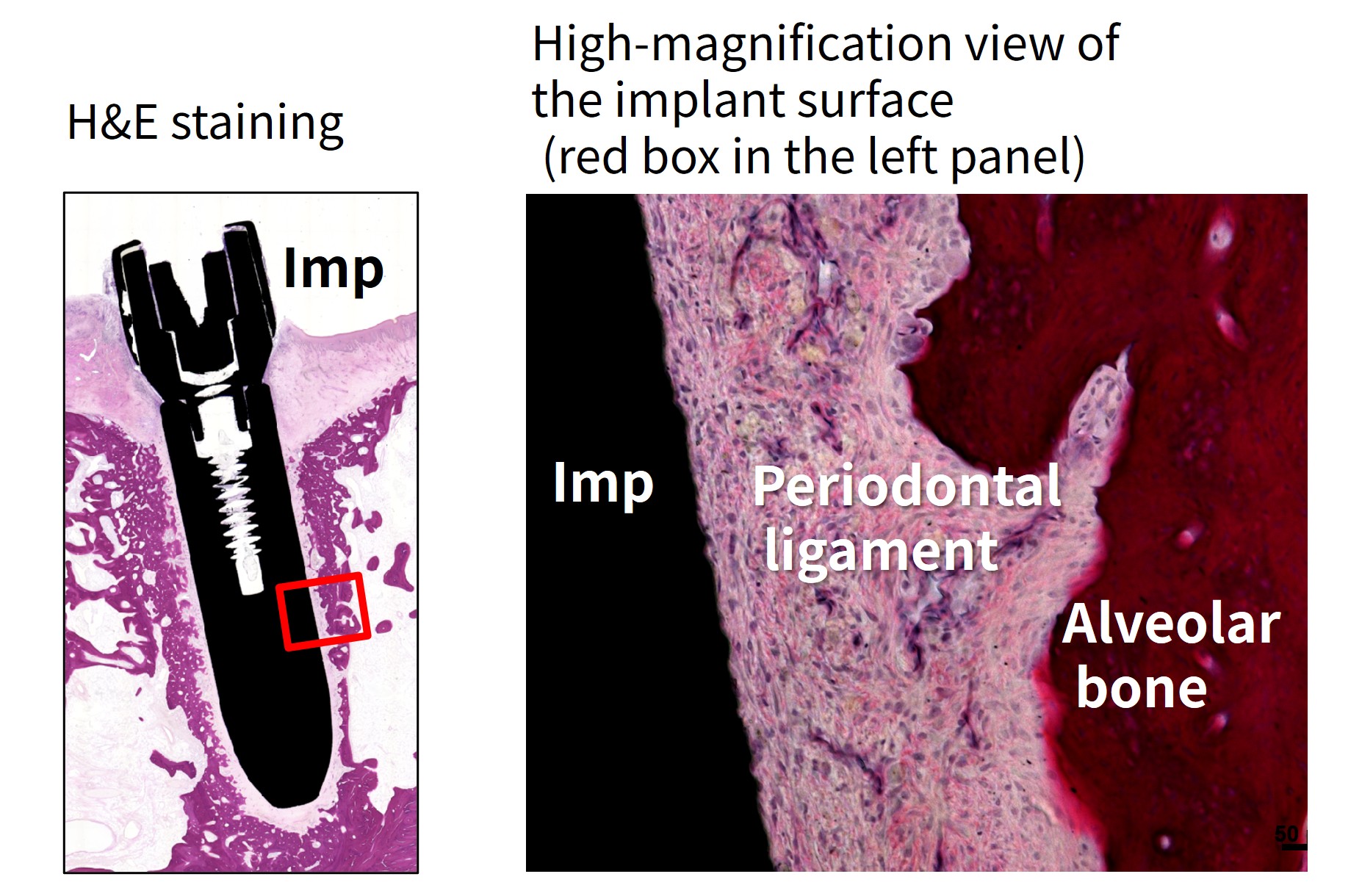
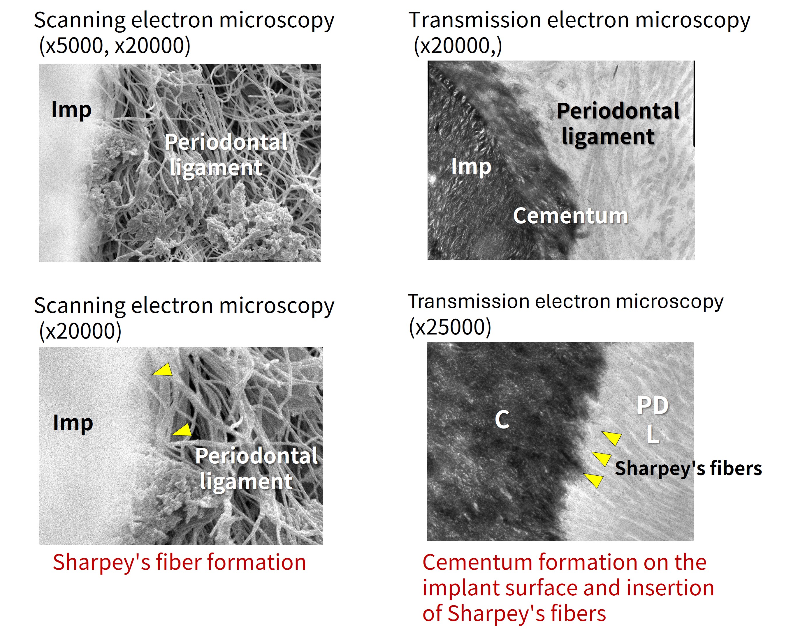
Histological analysis revealed a well-organized periodontal ligament structure comparable to that of a Natural tooth (Figure 4). Scanning electron microscopy demonstrated formation of Sharpey’s fibers within the periodontal ligament (Figure 5, left), while transmission electron microscopy showed lamellar Cementum formation on the implant surface with penetration of Sharpey’s fibers (Figure 5, right).
Functional biological evaluation using dynamic periodontal tissue testing (Periotest analysis) revealed that whereas an Osseointegrated implant typically exhibits negative values, the transplanted implant demonstrated a physiological mobility value of 2.9. This falls within the normal mobility range of canine Natural tooth (approximately 2–5), indicating successful engraftment of a bio-hybrid tooth mediated by the periodontal ligament.

These studies, progressing from mouse to canine models, experimentally demonstrated that periodontal ligament function can be conferred upon oral implants, establishing the feasibility of the bio-hybrid tooth concept.
However, numerous technological challenges still remained before clinical translation and application to human therapy could be achieved.